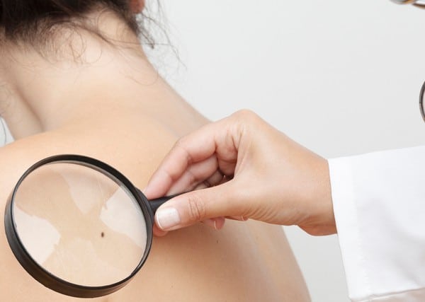
Dermoscopy is a vital tool in modern dermatology, enabling clinicians to evaluate skin lesions with greater accuracy than ever before. By providing a detailed view of the skin's surface, dermoscopy helps to identify various patterns that can distinguish between benign and malignant lesions. This guide will explore the Dermoscopy Mole Evaluation in Dubai , offering insights into their significance and application in everyday practice.
The Basics of Dermoscopy
What Is Dermoscopy?
Dermoscopy, also known as dermatoscopy or epiluminescence microscopy, is a non-invasive diagnostic technique used to examine the skin's surface. By magnifying the skin and illuminating it with polarized light, dermoscopy reveals subsurface structures and patterns that are invisible to the naked eye. This enhanced visibility is particularly useful in assessing pigmented lesions, aiding in the early detection of skin cancers such as melanoma.
Why Are Dermoscopic Patterns Important?
Dermoscopic patterns provide crucial information about the nature of a skin lesion. By recognizing and interpreting these patterns, clinicians can differentiate between benign and malignant lesions, leading to more accurate diagnoses and better patient outcomes. Understanding these patterns is essential for any clinician involved in skin assessments, as it reduces the reliance on biopsies and improves diagnostic confidence.
Key Dermoscopic Patterns and Their Significance
Reticular Pattern
The reticular pattern is one of the most common dermoscopic patterns observed in benign melanocytic lesions, such as common moles. It is characterized by a network of brown lines or "reticula" that form a honeycomb-like pattern. These lines correspond to the pigment distribution within the skin, with the darker areas representing increased melanin concentration.
- Significance: The presence of a regular, well-defined reticular pattern generally indicates a benign lesion. However, an irregular reticular pattern, particularly if it is broken or distorted, may raise suspicion of malignancy and warrants further investigation.
Globular Pattern
The globular pattern consists of multiple round or oval structures known as "globules." These globules are typically brown or black and vary in size. The pattern is often seen in benign nevi, particularly in children and adolescents, where it may represent a phase of mole evolution.
- Significance: A uniform globular pattern is usually benign, but irregularly sized or spaced globules, especially if they are clustered asymmetrically, can be a sign of dysplastic nevus or melanoma.
Homogeneous Pattern
The homogeneous pattern, also known as the structureless pattern, appears as a uniform area of pigmentation without any discernible internal structures. It is most commonly associated with benign lesions like seborrheic keratosis or hemangiomas.
- Significance: While a homogeneous pattern is often benign, it can also be seen in some melanomas, particularly in lesions with a blue-black coloration. Therefore, the presence of a homogeneous pattern should be interpreted cautiously, especially if other suspicious features are present.
Parallel Pattern
The parallel pattern is mainly observed on the palms and soles, where skin structures differ from other body areas. It consists of parallel lines running across the lesion, which can be classified into parallel furrow, parallel ridge, or lattice patterns based on their specific arrangement.
- Significance: On acral sites, the parallel furrow pattern is typically benign, whereas the parallel ridge pattern may be associated with acral lentiginous melanoma, a subtype of melanoma that occurs on the palms, soles, and under the nails.
Starburst Pattern
The starburst pattern is characterized by radial streaks or lines emanating from the center of the lesion, resembling the rays of a star. This pattern is often seen in pigmented lesions with active growth, such as Spitz nevi, which are typically benign but can be difficult to differentiate from melanoma.
- Significance: The presence of a starburst pattern, especially with uniform radial streaks, is usually benign. However, asymmetric or irregular streaks should raise suspicion for melanoma and may require further examination or biopsy.
Multicomponent Pattern
The multicomponent pattern refers to a combination of different dermoscopic patterns within a single lesion. For example, a lesion may exhibit areas of reticular, globular, and homogeneous patterns simultaneously. This complexity often complicates the interpretation and increases the challenge of making a definitive diagnosis.
- Significance: Multicomponent patterns are commonly associated with atypical or malignant lesions. The presence of multiple dermoscopic structures, particularly if they are irregular or asymmetrical, may suggest melanoma, and such lesions should be evaluated carefully.
Blue-White Veil
The blue-white veil is a dermoscopic feature that appears as a confluent area of blue or gray color with an overlying white "veil." This pattern is typically observed in malignant melanomas and is associated with significant alterations in the skin's structure, such as regression or the presence of dense pigmentation in the deeper layers.
- Significance: The blue-white veil is a strong indicator of malignancy, particularly in melanoma. Its presence should prompt immediate further investigation and, in most cases, a biopsy to confirm the diagnosis.
Practical Application of Dermoscopic Patterns
Pattern Analysis in Clinical Practice
Incorporating dermoscopic pattern analysis into clinical practice requires not only knowledge of the patterns themselves but also an understanding of their context within the lesion and the patient’s overall clinical picture. Clinicians should approach each lesion systematically, starting with an overall assessment of symmetry and border regularity, followed by a detailed examination of the internal patterns.
The Role of Algorithms in Pattern Recognition
Several dermoscopic algorithms, such as the ABCD rule, the seven-point checklist, and the chaos and clues algorithm, can aid in pattern recognition. These tools provide a structured approach to assessing lesions, helping clinicians identify suspicious features and decide when further action is needed.
Combining Dermoscopy with Other Diagnostic Tools
While dermoscopy is a powerful tool, it is most effective when combined with other diagnostic methods, such as patient history, clinical examination, and, when necessary, histopathological analysis. By integrating dermoscopy with these tools, clinicians can enhance their diagnostic accuracy and provide the best possible care for their patients.
Conclusion
Understanding and interpreting dermoscopic patterns is essential for clinicians involved in skin lesion assessment. By familiarizing themselves with key patterns such as reticular, globular, homogeneous, parallel, starburst, multicomponent, and blue-white veil, clinicians can improve their diagnostic capabilities and contribute to better patient outcomes. Dermoscopy, when used effectively, not only aids in the early detection of skin cancers but also reduces unnecessary biopsies, ensuring a more efficient and patient-centered approach to dermatologic care. As clinicians continue to refine their skills in dermoscopic pattern recognition, the benefits of this invaluable tool will only continue to grow.


Comments