Advanced imaging technologies have transformed how dental professionals diagnose and treat their patients. One such innovation is Cone Beam Computed Tomography (CBCT), a three-dimensional imaging technique that offers unparalleled insights into oral health. Unlike traditional two-dimensional X-rays, CBCT provides a comprehensive view of a patient’s dental anatomy, allowing for more accurate assessments and informed treatment decisions. This article delves into the five key benefits of a CBCT scan, elucidating its critical role in enhancing dental care.
Enhanced Diagnostic Accuracy
One of the foremost advantages of CBCT scans is their ability to deliver high-resolution images that significantly enhance diagnostic accuracy. Due to their two-dimensional nature, traditional X-rays can often obscure critical details, leading to potential misdiagnoses or overlooked conditions. In contrast, CBCT imaging provides a detailed three-dimensional perspective of the teeth, jaw, and surrounding structures, allowing dental professionals to identify issues such as impacted teeth, bone loss, and tumors with remarkable precision.
For instance, when evaluating a patient for dental implants, a CBCT scan reveals the precise anatomy of the jawbone, including its density and volume. This crucial information aids in more effectively planning the implant placement, ensuring better outcomes, and reducing the risk of complications. Visualizing the intricate relationships between teeth, roots, and adjacent structures is invaluable, empowering dental professionals to make more accurate diagnoses and tailor treatment plans accordingly.
Improved Treatment Planning
CBCT scans' role extends beyond diagnostics; they enhance treatment planning across various dental specialties. Whether it’s orthodontics, periodontics, or oral surgery, having access to detailed 3D images allows practitioners to devise more effective and individualized treatment strategies. For instance, orthodontists can assess the spatial relationships of teeth and roots, determining the most efficient path for tooth movement.
In oral surgery, CBCT scans provide essential information regarding the proximity of vital anatomical structures, such as nerves and sinuses, to the surgical site. This information is crucial in minimizing risks during tooth extractions or jaw surgeries. By utilizing CBCT scans in the treatment planning phase, dental professionals can significantly enhance the overall quality of care, ensuring that patients receive the most appropriate and effective treatments tailored to their unique needs.
Minimized Patient Discomfort and Radiation Exposure
A common concern among patients regarding dental imaging is radiation exposure. While traditional imaging techniques may require multiple X-rays to obtain the necessary information, a single CBCT scan can capture comprehensive data in one session. This efficiency minimizes the number of images taken and reduces patients' overall radiation exposure.
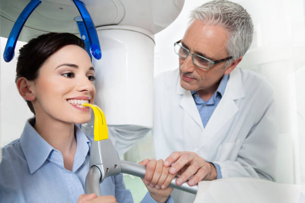
Moreover, CBCT scans are often more comfortable for patients than conventional imaging methods. The procedure typically involves the patient remaining stationary while the machine rotates around their head, capturing the necessary images. This streamlined process enhances patient comfort and contributes to a more efficient workflow in the dental practice, allowing for quicker diagnoses and treatments.
Facilitated Communication and Patient Education
Another significant benefit of CBCT scans is their ability to enhance communication between dental professionals and their patients. The visual nature of 3D imaging allows practitioners to share clear and comprehensive representations of a patient’s dental condition. This transparency fosters a better understanding of the diagnosis and proposed treatment plans, empowering patients to make informed decisions about their oral health.
For example, when discussing the need for a dental implant, a dentist can use CBCT images to show the patient the precise location of bone loss or the proximity of adjacent teeth and nerves. This visual aid not only demystifies the treatment process but also helps alleviate anxiety by providing patients with a clear understanding of what to expect. By facilitating open communication, CBCT scans play a crucial role in strengthening the dentist-patient relationship, ultimately improving treatment adherence and satisfaction.
Comprehensive Assessment for Diverse Dental Needs
The versatility of CBCT imaging makes it an invaluable tool for a wide array of dental applications. From endodontics to oral pathology, the comprehensive assessment capabilities of CBCT scans address diverse dental needs. In endodontics, for instance, the detailed visualization of root canal systems aids in diagnosing complex cases of infection or anatomical anomalies.
Similarly, oral pathologists can use CBCT scans to identify lesions, cysts, or tumors more accurately, allowing for timely and appropriate interventions. Assessing the full scope of a patient’s oral health through a single imaging modality streamlines workflows and enhances the overall quality of care provided. As dental practices evolve, integrating CBCT scans into routine assessments will likely become increasingly standard, reflecting the ongoing commitment to comprehensive patient care.
In summary, the incorporation of CBCT scans into dental practice represents a significant advancement in the field of dentistry. With their capacity to enhance diagnostic accuracy, improve treatment planning, minimize patient discomfort, facilitate communication, and provide comprehensive assessments, CBCT scans undeniably reshape the landscape of dental care. As technology evolves, embracing these innovations will be crucial for dental professionals striving to deliver their patients the highest standard of care. With the combination of detailed imaging and patient-centered approaches, the future of dentistry looks promising, ensuring better outcomes for all.


Comments